Brain PET Scan (Positron Emission Tomography) Q&A
Brain PET scans provide a rare window into how the brain functions and provide vital information for the diagnosis and treatment of a wide range of brain-related illnesses. The preparation and method for a PET scan are simple, with an emphasis on the patient’s comfort and imaging accuracy. At the iVIE MRI Screening Center, Dr. Kourosh Naini offers brain PET scans. For more information, contact us or book an appointment online. We are located at 11600 Washington Pl Suite 104A, Culver City, CA 90066.
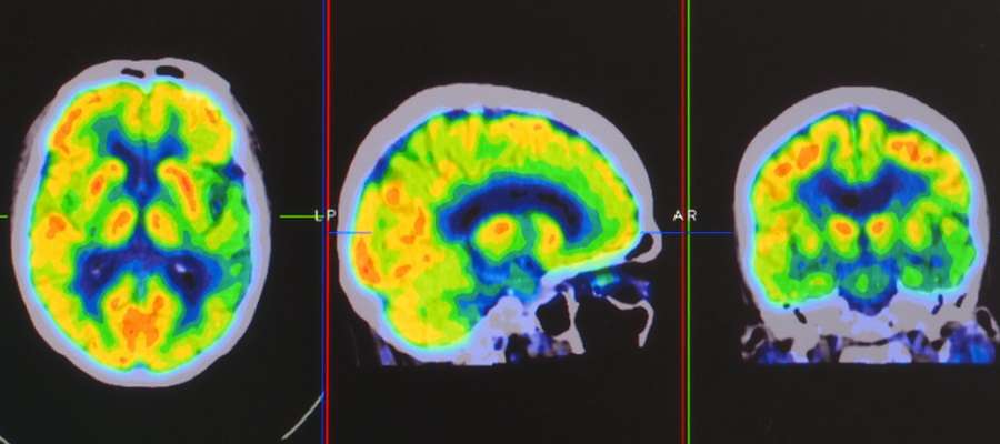
Table of Contents:
What is a brain PET scan?
Why is a brain PET scan performed?
How to prepare for a brain PET scan?
What happens during a brain PET/CT scan?
A brain Positron Emission Tomography (PET) scan is an imaging test that uses a radioactive substance, known as a tracer, to look for disease or injury in the brain. Unlike other imaging tests like MRI and CT scans that primarily reveal the structure of the brain and its surrounding tissues, a PET scan shows how the brain and its tissues are functioning.
This functionality aspect is crucial, as active areas of the brain utilize glucose at a higher rate than inactive areas. When highlighted under a PET scanner, it allows doctors to observe the brain’s workings and detect any abnormalities, providing detailed information on the size, shape, and function of the brain. PET scans are particularly effective in examining the brain’s chemical function, offering insights into organs and tissues at work.
Brain PET scans are frequently used to diagnose and assess the treatment of various conditions. They are essential in the treatment of patients with conditions affecting memory, movement, and other brain functions, including Alzheimer’s disease, epilepsy, stroke, and Parkinson’s disease.
PET scans can detect cancers and organs that are not functioning normally, like areas of the brain affected by Alzheimer’s disease or parts of the heart damaged by blocked blood vessels. Additionally, PET scans are combined with CT scans to provide more comprehensive details of both the function and structure of the brain for a more accurate diagnosis.
Preparing for a brain PET scan involves several steps to ensure accurate results. Instructions will be provided to patients by the specialists at the iVIE MRI Screening Clinic. Patients are generally advised to avoid strenuous exercise and physical activity for 24 hours before the appointment. It’s important to avoid caffeine and decaffeinated beverages for 24 hours before the exam.
Patients should stay well hydrated, drinking at least eight glasses of water, unless they are on fluid restriction. For six hours before the appointment, it’s recommended not to eat or drink anything other than water. Patients may be asked to change into a hospital gown and should wear comfortable clothing for the scan.
They should also leave valuables like jewelry at home to prevent misplacement. In preparation for the scan, an intravenous (IV) line is placed into a vein, through which a radioactive tracer is injected. It takes about 30 to 90 minutes for the tracer to travel throughout the body, during which time the patient needs to remain still and relaxed.
During a brain PET/CT scan, patients lie down on a table, which moves them through the scanner. The machine has a larger opening than an MRI scanner and does not produce any sound. The PET scanner detects diseased cells that absorb large amounts of the radiotracer, indicating potential health problems.
The camera used in the scan detects emissions from the injected radiopharmaceutical, and the computer attached to the camera creates two- and three-dimensional images of the examined area.
The areas where the injected radiopharmaceutical accumulates, such as fast-growing cancer cells, appear ‘brighter’ than normal tissues on the images. This combination of PET and CT scanners allows the nuclear medicine specialist to combine structural information from the CT scan with the PET’s functional information, enhancing the accuracy of the test.
After the test, patients can usually resume their day as usual, unless advised otherwise. They are encouraged to drink plenty of fluids to help flush the tracer from their body.
A PET scan procedure typically takes about an hour to complete and generally does not require an overnight hospital stay. If a contrast medium is used, it can either be ingested or injected through the IV. The scanning process involves the table sliding through the scanner, with the patient needing to remain still, sometimes holding their breath to avoid blurry images.
The technologist might adjust the table’s position to obtain images from different angles. The entire process, from the initial preparation to the completion of the scan, takes about two hours.
Brain PET scans are available at the iVIE MRI Screening Center. For more information, contact us or book an appointment online. We are located at 11600 Washington Pl Suite 104A, Culver City, CA 90066. We serve patients from Culver City Los Angeles CA, Downtown LA, Beverly Hills CA, Marina del Rey CA, Venice CA, Playa Vista CA, Santa Monica CA, and surrounding areas.
Additional Services You May Need
▸ Dementia Screening
▸ Aneurysm Screening
▸ Spine MRI
▸ Whole Body MRI Screening
▸ MRI Brain Screening
▸ Brain PET
▸ Work/Sport Spine Injury Diagnosis
▸ Work/Sport Brain Injury Diagnosis
▸ Whole Body PET For Cancer
▸ MRA Brain Screening

